
Research Summary: Comparative morphology of the talus bone of Plio-Pleistocene vertebrates
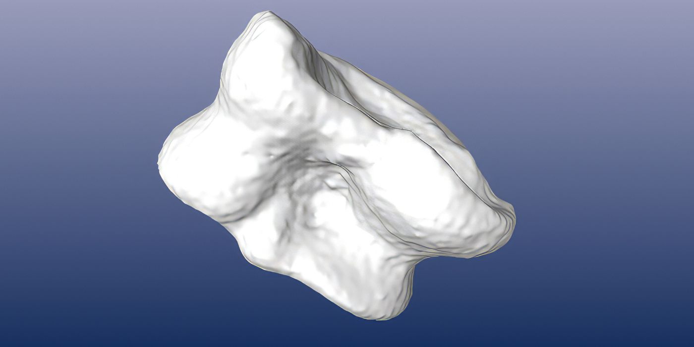
My research hypothesizes that with an effective "whole 3D surface ordination" technique statistically significant variation in vertebrate morphologies can be quantified even from small, isolated and feature-poor post-cranial bone elements to permit the consistent identification of genera respectively species. "Whole 3D surface ordination" is my working title for the core of this research effort: to find a new technique for using regular geometric morphometrics without resorting at all to anatomically homologous landmarks. Even the latest attempts to incorporate entire 3D surfaces in geometric morphometrics, such as Eigensurface Analysis (Polly and MacLeod), use at some step user-specified landmarks. These may not be placeable either because the bone is fragmentary or indeed does not have such features. Hence, I've chosen to test out the hypothesis with fossilized talus bones (pl. astragali, sg. astragalus) of vertebrates mainly from the Bovidae family from the limited geographical and temporal range of the African Pliocene and early Pleistocene epochs (5.3 to 1.4 million years into the deep past). Astragali have few anatomical features on which landmark points can be consistently placed but are central to locomotion and are often found during surface fossil surveys because of their compact form.
This research contributes to applied geometric morphometrics and impacts the broader fields of paleoecology and evolutionary development biology. By being able to identify taxa with greater precision and confidence, both the range and the quality of specimen data for the purpose of reconstructing the environment in which human ancestors lived, will be greatly expanded. By intensely studying a single structure, which does have an important functional role in locomotion, we gain additional insights into how environmental changes and pressures are revealed in evolved morphologies. In designing an efficient workflow to quantify 3D geometric variation in fossilized bone structures over a large set of specimens, and doing so in such a way that other researchers can reproduce the results, the potential of the technique would be demonstrated for use with other similarly landmark-poor post-cranial body elements.
This research builds on the comprehensive linear measurement set on astragali from yet to be published work by René Bobe et al (George Washington University, USA). That data set has been made available. I will work with many of the same specimens but will quantify their entire 3D surface morphologies rather than using simple linear measurements or derived linear ratios.
The astragalus is seen as a key skeletal element as it transfers the weight of the main body distally to the tarsals. It has been shown that astragali will significantly differ in their morphology as conditioned by the taxa's general form of locomotion (e.g. fast running on open plains, hiding in wooded areas, ...). Previous work, however, was motivated by connecting extant species with known preferred habitats with Plio-Pleistocene fauna by way of astragali data for the purpose of paleoenvironmental reconstruction. This research is, by extension, instead interested in identifying the specific genera respectively species.
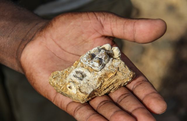
Astragali are often found during surface fossil surveys. A higher probability of discovery can be presumed. Astragali are compact and of relative small size. Compared to long bones, they may be less likely to get trampled by game or to be broken apart by geomorphological processes over evolutionary periods of time. Hence, being able to securely assign astragali to their taxon will aid future fossil surveys.
Data collection:
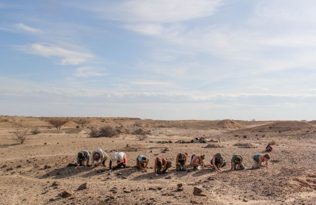
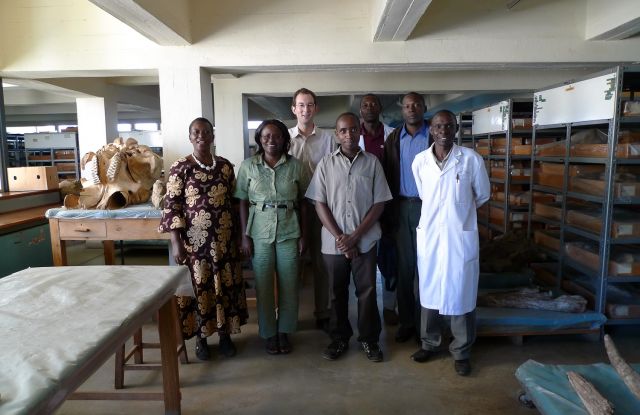
Data will be collected from the astragalus specimens within the extensive paleontological collection of the National Museums of Kenya in Nairobi, including the large set of astragali from this year's Koobi Fora surveys to which I contributed. Virtual replica of each of the astragali will be produced by close-range photogrammetry and/or 3D laser scanning for subsequent data reduction.
Data reduction and analysis:
The taxa assignment of astragali will not be assumed a priori. Instead, cluster analysis will be performed on the 3D surface measurement data to discover morphologically quasi-homogenous groups. The research will determine which of the various statistical clustering methods is the most effective and appropriate. Clustering operations on high-dimensional data has been recognized as a particular challenge in the data reduction. Expectations are that for the certain genera and, possibly, species, clusters will be identifiable.
Prerequisites:
- Continued access to the fossil collection for the next few months would need to be granted. By start of this research, the investigator will already have worked for over a month with the collection.
- Reasonably easy access to the relevant citation databases for a comprehensive review of the existing literature in the field would be required.
Main tasks and recognized challenges:
- Set up 3D scanning station and design 3D point cloud post-processing. The key challenge will be the production of noise-limited 3D mesh surfaces.
- Set up imaging station, calibrate camera and practice the imaging workflow for close-range photogrammetry. The key challenge will be to control lighting and construct noise-limited 3D mesh surfaces from individual frames.
- Search citation databases for additional literature not only on geometric morphometrics but on the wider field of 3D surface quantification, both computational methodologies and practical applications. The key challenge will be to indeed find and review all relevant literature.
- Use regular geometric morphometrics, including Procrustes superimposition and Principal Component Analysis. Try out alternative ordination and clustering methodologies. The key challenge is to find a technique to do so with entire 3D surfaces without resorting to homologous landmarks.
- Write up final report
Collaboration:
Professor René Bobe has provided the initial data set. The principal investigator would be delighted if Kenyan research associates/students interested in comparative morphology would choose to join this research, from the start or at a later stage.
Funding and equipment:
The principal investigator is self-funded. Equipment and software applications for 3D laser scanning, 2D imaging for 3D reconstruction, linear measurements and Procrustes and Principal Component Analysis are available.
Additional benefits of the research:
The optimized workflow for close-range photogrammetry and the 3D models and imagery will be made available to the National Museums of Kenya. It may be interesting to explore their use for public display and education.
Temporary contact information for the principal investigator:
Postal address: Marcel Bigger, Paleontology Division, Department of Earth Sciences, National Museums of Kenya, Museum Hill, 00100 Nairobi, Kenya
Kenyan mobile telephone number: 0729 810 146
Background:
During a paleoanthropological field expedition to the Lake Turkana region near the Kenyan-Ethiopian border and subsequent curatory work at the National Museums of Kenya, the principal investigator was intrigued that fossil specimens were assigned to taxa mostly by way of visual inspection and comparison with curated samples. Additionally, it appeared that the discipline of geometric morphometrics has focused on specimens respectively bone elements with easily definable landmarks. This was taken as a challenge to do geometric morphometrics on a bone element distinguished by few point landmarks, the astragalus.


